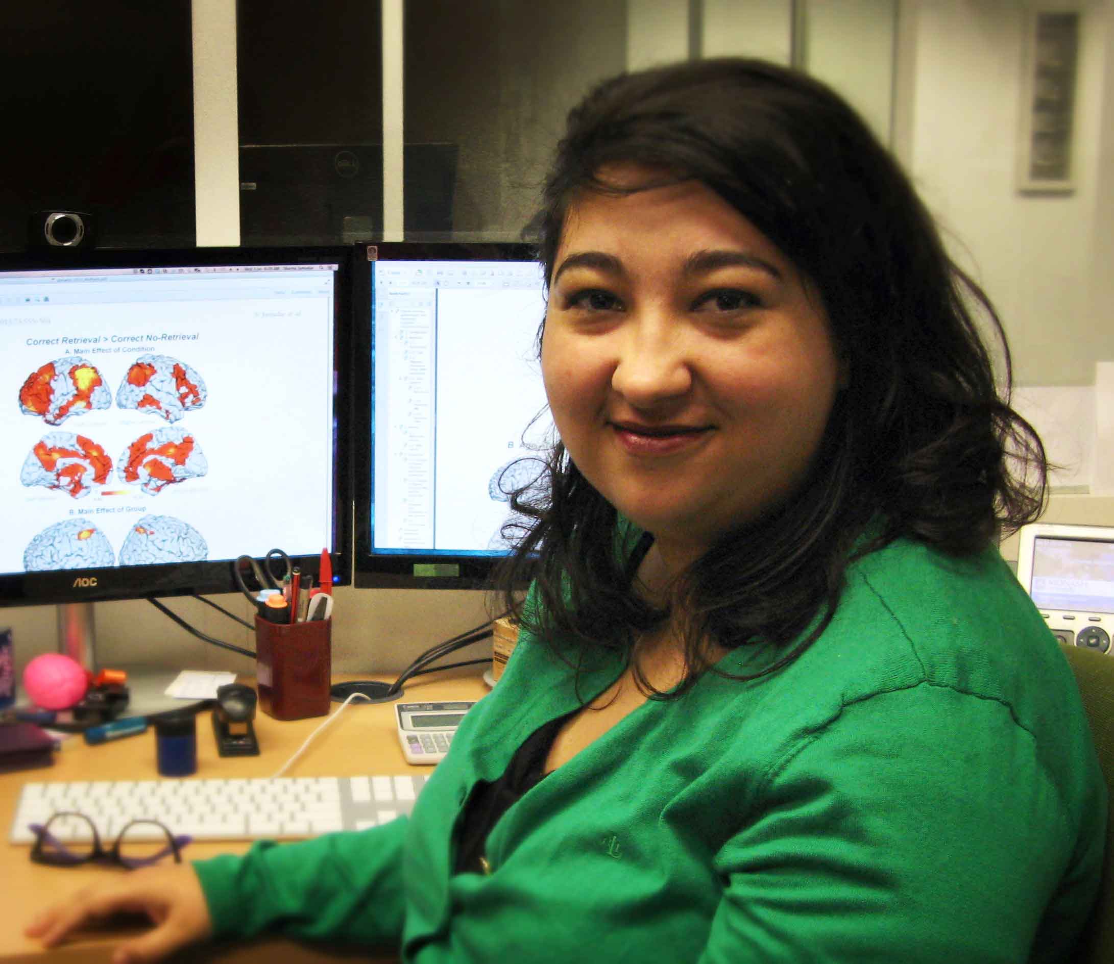
Dr Sharna Jamadar is one of the first researchers to complete our console operator training. She undertook the training at Monash Biomedical Imaging (MBI) and finished earlier in 2014. In late May we had a chat with her about the training. Here is what she had to say:
Hi Sharna, thanks for taking the time for this interview, we appreciate it.
1. So, tell us about yourself?
Since the beginning of my research career, I have been interested in cognitive control processes – i.e. those processes that allow us to flexibly and dynamically change our behaviour to respond appropriately to changing environmental circumstances. I use multiple cognitive neuroscience techniques to study cognitive control, particularly electroencephalography/event-related potentials (EEG/ERPs) and functional magnetic resonance imaging (fMRI). I find that combining information across multiple neuroimaging techniques is a powerful way to understand the underlying cognitive processes that contribute to the overt behaviour.
I completed my PhD in 2010 on Psychology/Cognitive Neuroscience at the University of Newcastle. The broad goal of my PhD was to use multiple neuroscience and neuroimaging techniques (i.e. behavioural, EEG/ERP and fMRI measures) to examine cognitive control using the task-switching paradigm. Following my graduation, I took up a Postdoctoral Fellowship at the Institute of Living in Hartford, CT USA. It was a highly cross-disciplinary environment and I extended my skill base into imaging genetics and structural imaging in the study of cognition and genetic markers in schizophrenia, ageing, memory and genetic susceptibility to alcoholism. Since 2012 I have been a Research Fellow at Monash Biomedical Imaging (MBI) and School of Psychological Sciences (SPS) at Monash University. A large focus of my time at Monash has been the development of cognitive neuroscience research facilities and research at MBI. My research focus has continued to be on the use of multiple neuroscience techniques in the study of higher-order cognition, this time combining oculomotor (eye-tracking) outcomes with fMRI.
2. In what ways do you use MRI?
As a cognitive neuroscientist, I am primarily interested in using neuroimaging techniques to understand the underlying neural processes involved in behaviour, and how differences in brain function can account for differences in behaviours observed between groups and individuals. The primary focus of my MRI work is with functional brain MRI, in particular task-based fMRI but also resting-state fMRI. I also have an interest in structural brain MRI and how neuroanatomical differences between groups and individuals can account for variation in behaviour.
3. Why is learning to operate the MRI console of value to you?
Being able to operate the MRI console gives me greater flexibility as a researcher. Quite often it can be difficult to organise participants around a typical Monday-Friday 9-5pm schedule – not only because during this time of high-demand it can be difficult to work around other researchers using the MRI scanner, but also because many participants find it difficult to take time out on a weekday to attend an appointment that sometimes requires a 2-3hr time commitment. So, by being able to operate the scanner myself, I can offer my participants greater flexibility.
In addition, being able to operate the MRI console gives me greater control over the data quality and outcomes. For example, if I’m interested in preferentially imaging one brain area over another, then I am in full control over the alignment of the field of view to do so.
4. Tell us about your experiences of the process.
I was very excited when offered the chance to learn how to operate the MRI scanner as it was something that I had wanted to do for a long time. My primary motivation was for the increased level of flexibility and control it would give me to be able to acquire my own fMRI data, just as I have always acquired other forms of data like EEG. Before I started, I expected that I’d basically just be shown how to use the console software for the types of imaging protocols that I usually use (e.g. BOLD-fMRI, MPRAGE) and then I’d scan a whole heap of brains until I’d completed my hours. But the training protocol offered so much more than that, and I think it has helped me become a better researcher. My primary training mentor, Senior Radiographer Richard McIntyre, took a very didactic approach and taught me a lot about MRI as a technology. As a cognitive neuroscientist, learning about MRI technology was something that I picked up a bit by ‘osmosis’, and a bit from reading in my spare time. Richard and the other radiographers taught me a lot more about parameters that I had only a vague notion of, and some that I hadn’t even encountered before.
Also, the requirement to scan phantoms, ex-vivo samples and animal subjects for 20hrs was something that I initially thought would be tedious and laborious with little benefit. But in fact, I found that I probably learned more during this part of the training than in any of the other components of the training. Scanning the phantoms gave me the opportunity to play around with different parameters to see the effects on the resulting images, and scanning the ex-vivo samples and animal subjects gave me the opportunity to try new techniques like MR spectroscopy. This part of the training also made me realise just how human neuro-centric my view of MR images are, and that my sense of space is completely off when looking at anything other than a human brain!
As one of the first to complete the training at MBI, lots of people have asked me about how I’ve felt about the training protocol and the time commitment required to complete it. The protocol itself is very demanding when compared to what is required to operate an MR scanner in a research setting in other countries, but I think that the benefit of undertaking the full protocol exceeds any costs associated with time and effort. My advice to others about to undertake the training is to make the most of all the aspects of the protocol, even the parts that might seem tangential to your research needs in the beginning. And most importantly, make the most of your time with the radiographer mentoring you, I learned so much just from my interactions with them over the course of the training!
At the completion of the protocol, I feel confident that I can complete a human MR scanning session from the very start to the end, from starting up the scanner, loading protocols, screening participants, participant positioning, running the scans, troubleshooting, data archiving and scanner shut-down. I am now in a position of control over the data acquisition and its quality, and have much more flexibility in when I can schedule my participants’ appointments.
5. Thanks for your time Sharna. I’m glad you found the training so valuable. Perhaps you can send us a copy of the first paper where you are an author and an MRI console operator?
You are welcome. I’m very happy that Australian researchers now have the opportunity to operate the MR console at research institutions – my friends and colleagues from other Australian institutions who have not yet implemented the protocol are very jealous! I’ll definitely send through a copy of the first paper, and I’ll also send you some of the images I have taken.
Thanks to VBIC and MBI for the opportunity.


0 Comments